
610-459-9646
sales@sciotex.com
ENGINEERING GUIDE
Light Sheet Microscopy: How It Works, Types and Instrument Control
How light sheet fluorescence microscopes image living samples in 3D, how SPIM, multiview, Bessel, lattice and adaptive optics systems differ, and what it takes to control one.
Last updated October 2026 · Written by Sciotex Engineering
DEFINITION
What is light sheet microscopy?
Light sheet microscopy, also called light sheet fluorescence microscopy (LSFM) or selective plane illumination microscopy (SPIM), illuminates a sample from the side with a thin sheet of laser light and records the fluorescence with a camera on a perpendicular detection axis. Only the plane being imaged is lit, so each image is optically sectioned and the rest of the sample is spared. Stepping through the specimen builds a 3D volume; repeating that builds a 3D movie.
The names overlap. SPIM was the original name for the technique, LSFM is the umbrella term, and light sheet microscope covers both commercial systems and instruments built in research labs. Variants such as multiview, Bessel beam and lattice light sheet microscopes change how the sheet is formed or how many views are recorded, but the principle is the same.
This guide explains how light sheet microscopy works, the main types, how it compares with confocal and two-photon microscopy, and the part most introductions skip: how these instruments are controlled. Sciotex has developed custom control software for light sheet, lattice light sheet and adaptive optics microscopes, including instruments at HHMI’s Janelia Research Campus.
HOW IT WORKS
How light sheet microscopy works
A light sheet microscope separates illumination and detection onto two perpendicular axes. That one design choice is what makes it fast and gentle.
Because a whole plane is captured in one camera exposure, rather than point by point as in a confocal, light sheet systems can record hundreds of planes per second. And because only the imaged plane is lit, the sample receives far less total light, so living embryos and cells can be imaged for hours or days. That gives light sheet microscopy six practical advantages:
Optical sectioning
Only the focal plane is illuminated, so out-of-focus blur is largely avoided at the source.
Low phototoxicity
Far less light reaches the sample, keeping live specimens healthy over long experiments.
Speed
Whole planes are captured per camera frame, enabling fast volumetric imaging.
Large samples
Embryos, organoids, whole brains and cleared organs can be imaged in 3D.
Long time-lapse
Low bleaching makes multi-hour and multi-day recordings practical.
Quantitative 4D data
Volumes over time support cell tracking, lineage tracing and 4D analysis.
TYPES
Types of light sheet microscopes
Light sheet microscopes differ mainly in how the sheet is formed and how many views they record. Further down the table, systems generally trade simplicity for resolution, coverage or depth.
| Type | How the sheet is made | Strengths | Typical use |
|---|---|---|---|
| SPIM (static sheet) | A cylindrical lens forms a fixed sheet | Simple, robust, fast | Embryos and larger specimens |
| Scanned light sheet | A galvo scans a thin beam to build the sheet | Uniform sheet, adjustable thickness | General live imaging |
| Multiview light sheet | Two or more illumination and detection arms record several views at once | Even coverage of large, scattering specimens | Whole embryos, developmental biology |
| Bessel beam light sheet | A scanned Bessel beam gives a thin, extended sheet | Thinner sheet, better axial resolution | Single cells and subcellular imaging |
| Lattice light sheet | A spatial light modulator shapes a 2D optical lattice into an ultrathin sheet | Very high resolution with low phototoxicity | Subcellular dynamics in living cells |
| Adaptive optics light sheet | Deformable mirrors or SLMs correct wavefront aberrations | Sharp images deep inside tissue | Cells within living organisms |
| Cleared tissue light sheet | Large-field, long working distance optics for transparent samples | Whole organs at cellular resolution | Neuroscience and anatomy |
Swipe the table to see all columns →
Sciotex has developed control software across this range, including multiview, Bessel beam, lattice and adaptive optics systems. For more depth, see lattice light-sheet microscopy and adaptive optics microscopy.
COMPONENTS
Light sheet microscope components
Every light sheet microscope, from a teaching setup to a lattice system, is built from four groups of parts. Getting them to work together is mostly a timing and software problem.
Illumination: lasers and sheet forming
Lasers at one or more wavelengths, switched by acousto-optic modulators or Pockels cells. The sheet is made by a cylindrical lens, a galvanometer mirror scanning a beam, or a spatial light modulator for Bessel and lattice sheets.
Detection: objective, filters and camera
A detection objective at 90 degrees to the sheet, emission filters and fast scientific cameras, usually sCMOS. See sCMOS vs EMCCD cameras.
Sample positioning and motion
Piezo stages or objective scanners step through the sample, while motorized stages and rotation mounts position it and record extra views.
Control, acquisition and data
DAQ or FPGA hardware that times every device, acquisition software that runs the experiment, and storage fast enough for gigabytes per second. See data acquisition.
INSTRUMENT CONTROL
Synchronization: the hardest part of a light sheet microscope
The optics get the attention, but most of the engineering in a working light sheet microscope is in keeping every device in step. One volume can involve hundreds of camera exposures, each matched to a galvo position, a piezo step and a laser pulse, repeated for hours.
COMPARISON
Light sheet vs confocal vs two-photon microscopy
All three image fluorescent samples in 3D, but they spend light very differently. Confocal and two-photon microscopes scan a focused point through the sample; light sheet microscopes illuminate a whole plane at once.
Choose light sheet when…
- You need fast 3D imaging of living samples
- Phototoxicity or bleaching limits how long you can image
- The sample is large: embryos, organoids or cleared organs
- You need long time-lapse recordings at high time resolution
- The sample can be mounted for side illumination
Choose confocal or two-photon when…
Confocal microscopy remains the flexible workhorse for fixed samples, thin specimens and standard slides, where mounting a sample for a light sheet would be awkward. Two-photon microscopy reaches deeper into scattering tissue, such as the brain of a living animal, because its infrared excitation scatters less. Many labs use all three, and adaptive optics can extend both two-photon and light sheet imaging deeper.
APPLICATIONS
Light sheet microscopy applications
Speed, gentleness and 3D coverage have made light sheet microscopy a standard tool wherever living specimens must be watched in three dimensions over time.
1Developmental biology
Imaging whole zebrafish, fruit fly and other embryos for hours or days as they develop, often with multiview systems that capture the entire specimen.
2Cell biology
Following organelles, the cytoskeleton and membranes inside living cells at high resolution, where Bessel and lattice light sheets excel.
3Neuroscience
Whole-brain functional imaging in small model organisms, and cellular-resolution maps of cleared brains.
4Cleared tissue and organs
Imaging whole organs and tissue blocks made transparent by clearing, at cellular resolution in 3D.
5Organoids and 3D cultures
Watching organoids and spheroids grow and respond to drugs, with less light damage than point-scanning methods.
6Deep-tissue imaging
Combining light sheets with adaptive optics to image cells inside living organisms, where tissue would otherwise blur the image.
BUILDING ONE
Building or upgrading a light sheet microscope: the software side
Commercial light sheet systems cover many needs, but research groups often build or modify instruments for a specific experiment. The optics are only half the job; these software and control steps decide whether a custom light sheet microscope works day to day. See also microscope hardware synchronization.
1. Define the experiment
Sample, labels, volume size, speed and duration set every other requirement.
2. Plan the timing architecture
Choose DAQ, FPGA or real-time hardware that can drive galvos, piezos, lasers and cameras from one clock.
3. Synchronize the hardware
Generate hardware-timed waveforms and camera triggers so every exposure matches the sheet position.
4. Build the acquisition pipeline
Stream multi-camera data to disk without dropped frames, with metadata for every plane.
5. Add calibration and alignment tools
Sheet-to-focal-plane alignment, galvo and piezo calibration, and checks users can run themselves.
6. Make it usable and supported
Interfaces scientists can run without the developer, and support as experiments change.
DATA RATES
Light sheet data: planning for gigabytes per second
Light sheet microscopes are some of the most data-hungry instruments in a lab. Estimating the data rate early decides the cameras, computers, storage and network the system needs.
Worked example. A 2048 × 2048 pixel sCMOS camera at 16 bits (2 bytes per pixel) produces 2048 × 2048 × 2 ≈ 8.4 MB per frame. At 100 frames per second that is about 839 MB/s; with two cameras, about 1.7 GB/s, or roughly 6 TB per hour of continuous imaging.
That is why acquisition software for light sheet systems is built around streaming, compression and fast storage, and why GPU and FPGA processing are often used to reduce data on the fly. Sciotex builds distributed multi-camera acquisition and GPU and FPGA image processing for exactly these loads.
REAL-WORLD EXAMPLES
Microscope and imaging systems Sciotex has worked on
Sciotex has developed more than 30 custom microscope software applications for over 15 research institutions. These are a few of the instruments and imaging systems.

Multiview light sheet microscope
Control software for a microscope with multiple illumination and detection arms, recording high-resolution time series of living samples.
View example →

Adaptive optics microscope
LabVIEW control of a custom two-photon microscope that corrects tissue aberrations with a spatial light modulator.
View example →
SLM phase pattern algorithm
GPU-accelerated phase patterns for spatial light modulators, for selective illumination with real-time feedback.
View example →
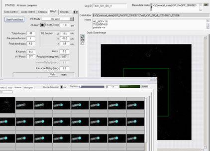
Confocal microscope
A confocal microscope with two-photon fluorescence capability, developed with University of Pennsylvania researchers.
View example →

Birefringence imager
Automated imaging of birefringent materials in 96-well plates with XY stage and dual polarizer control.
View example →

CellSpotter cell detection
Collects, analyzes and archives microscope images of blood samples to detect circulating cancer cells.
View example →
KEEP LEARNING
Microscopy articles
Lattice Light-Sheet Microscopy: How It Works
How optical lattices create ultrathin light sheets for subcellular live imaging.
Read article →
Adaptive Optics Microscopy
How wavefront correction keeps images sharp deep inside tissue.
Read article →
sCMOS vs EMCCD Cameras for Microscopy
Which scientific camera fits light sheet, confocal and low-light imaging.
Read article →
Microscope Hardware Synchronization
Timing galvos, piezos, lasers and cameras with DAQ and FPGA hardware.
Read article →
Laboratory Automation Software
Custom vs off-the-shelf software for automated lab instruments.
Read article →
AI in Biomedical Imaging Analysis
Where AI helps analyze microscopy and biomedical image data.
Read article →
FAQ
Light sheet microscopy FAQs
What is light sheet microscopy?
Light sheet microscopy, also called light sheet fluorescence microscopy (LSFM) or SPIM, lights a sample from the side with a thin sheet of laser light and images the fluorescence from a perpendicular axis. Only the imaged plane is illuminated, giving fast, optically sectioned 3D imaging with low phototoxicity.
What is the difference between light sheet and confocal microscopy?
A confocal microscope scans a focused point through the sample and uses a pinhole to reject out-of-focus light, so the whole depth is illuminated for every plane. A light sheet microscope illuminates only the plane being imaged and captures it in one camera frame, making it much faster and gentler for live 3D imaging.
What is SPIM microscopy?
SPIM, selective plane illumination microscopy, was the original name for light sheet microscopy, using a cylindrical lens to form a static sheet. Today SPIM and LSFM are often used interchangeably.
What is lattice light-sheet microscopy?
Lattice light-sheet microscopy uses a spatial light modulator to shape a two-dimensional optical lattice into an ultrathin light sheet, enabling high-resolution 3D imaging of living cells with very low phototoxicity. See lattice light-sheet microscopy.
What are the advantages of light sheet microscopy?
Optical sectioning at the source, low photobleaching and phototoxicity, high volumetric imaging speed, the ability to image large specimens such as embryos and cleared organs, and long time-lapse recordings.
What are the limitations of light sheet microscopy?
Samples must be mounted so they can be lit from the side, absorbing structures can cast shadows or stripes, scattering degrades the sheet in thick tissue, and the data volumes are very large. Multiview designs, adaptive optics and good acquisition software address many of these.
What camera is used for light sheet microscopy?
Most light sheet microscopes use sCMOS cameras for their speed, large sensors and low read noise. EMCCD cameras are still used for the lowest-light applications. See sCMOS vs EMCCD cameras.
What software controls a light sheet microscope?
Commercial systems ship with vendor software, and open-source tools are widely used. Custom and research instruments usually need custom control software that synchronizes the hardware and streams data, which is what Sciotex develops as custom microscope software.
Building or upgrading a microscope?
Sciotex has developed more than 30 custom microscope software applications for over 15 research institutions, including control software for light sheet, lattice light sheet and adaptive optics microscopes. A Sciotex engineer co-authored the 2014 Science paper that introduced lattice light-sheet microscopy.

© Copyright 2026 by Coleman Technologies, Inc. DBA Sciotex