Adaptive Optics Microscope Software
Adaptive Optics Microscope
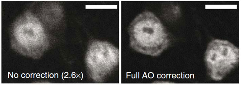
In biological imaging, the object of interest is not always in plain sight. This is especially true when trying to image cells buried deep within tissues. The tissues covering the cells tend to bend and scatter the light passing through them, preventing sharp images of the cells within. To overcome this, researchers at the Howard Hughes Medical Institute thought of a novel way to use adaptive optics.
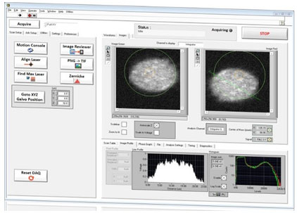
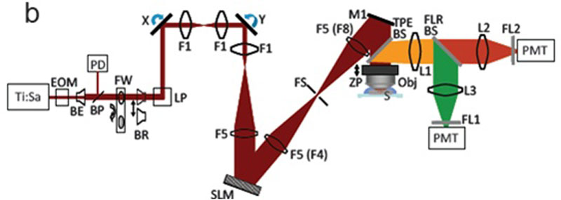
Adaptive optics is a technique used to correct for imaging aberrations. It is most famously known in astronomy where ground-based telescopes correct for how the atmosphere tends to bend and scatter starlight. In the case of biological microscopy, the technique can be adapted to correct for the biological aberrations. Here many images are initially taken to characterize the aberrations that each light path through the sample experiences. A spatial light modulator is then used to steer the parts of the light beam back into sharp focus. To develop the instrument control program to carry out this technique, the researchers worked with Dr. Daniel Milkie of Sciotex to build the LabVIEW program that would run the 2-photon microscope, analyze the images, and optimize the adaptive optics to correct the aberrations. This success of this work was recently published in Nature Methods. Nature Methods Article Abstract

Features
- LabVIEW control of a custom built two-photon microscope with intuitive user interfaces
- Multiple acquisition modes including single scans, Z-stacks, calibration routines, and adaptive optics scans
- Online reviewing and analysis suites, including image profiling, image statistics, Zernike deconvolution of aberrations
- Synchronized control of external hardware including : 1,920×1,080 pixel spatial light modulator, Pockel Cell, filter wheel, Galvo mirrors, beam reducers, and motion-stage elements
- Simultaneous high-speed DAQ input and waveform output using RTSI bus and multiple NI-DAQ cards
- Real-time adjustment of experimental parameters throughout the acquisition
- Multi-thread and multi-processor technologies to optimize system performance


Need a Consultation?
Save Time and Money with an Engineered Solution from Sciotex
© Copyright 2023 by Coleman Technologies, Inc. DBA Sciotex
